Macular Degeneration (AMD) Care in Chattanooga, Knoxville, and the Tri-Cities Region
Macular degeneration causes a gradual deterioration of the retina, affecting central vision while leaving peripheral vision intact. Commonly known as age-related macular degeneration (AMD), it is the leading cause of irreversible vision impairment in the United States, particularly among those over the age of 50. Southeastern Retina Associates offers advanced surgical and nonsurgical treatments to delay or halt AMD progression and preserve existing vision.
What is Age-Related Macular Degeneration?
Situated at the back of the eye, the retina is an ultrathin, light-sensitive tissue layer, about 0.5 mm thick. Through a process called transduction, the retina captures incoming light, converting it into electrical signals that travel along the optic nerve to the brain, transforming it into viewable images.
AMD slowly damages the central part of the retina, known as the macula, affecting the crucial photoreceptor cells responsible for advanced central vision tasks. Although AMD is incurable, proactive eye care can play a crucial role in preserving eye health and minimizing the impact of age-related changes on vision.
AMD is a progressive condition that is categorized into two subtypes: dry and wet.
Dry AMD
Dry AMD is the earlier, more common stage of macular degeneration. Typically, dry AMD is not destructive or painful; in some cases, it may even have very little impact on vision. However, if you do experience vision issues or loss, you may experience it in one or both eyes, gradually worsening. In most cases, this stage is identified during a normal dilated eye exam. Once diagnosed, many patients do well without treatment, although regular monitoring will be needed. However, you must alert your retina specialist immediately as soon as you experience any vision changes, as this may signal the development of more advanced stages.
The most severe form of dry AMD is geographic atrophy, a chronic and progressive eye condition that gradually causes the macula’s cells to deteriorate, leading to irreversible central vision loss.
Wet AMD
Affecting about 10% of AMD patients, wet AMD is much more serious. Also called exudative AMD, it’s characterized by choroidal neovascularization (CNV) – a process characterized by the growth of unusual blood vessels in the choroid and emerging beneath the retina. As these new vessels are fragile and easily broken, blood and other fluids can seep into the macular area. Symptoms of wet AMD can include central vision blurriness, distortions, and worsening vision loss. Also, compared to dry AMD, vision loss is much more rapid and noticeable with wet AMD.
Age-Related Macular Degeneration Risk Factors
With both dry and wet AMD, being over 50 years old is the leading risk factor. Certain disorders and lifestyle factors may increase your likelihood of its development, including:
- A family history of AMD
- Underlying conditions, including diabetes, high blood pressure, or high cholesterol
- Having light-colored irises (the colored eye part surrounding the pupil that regulates incoming light levels)
- Obesity
- Smoking (all types)
- A poor diet, such as one with high saturated fats
- Long-term, unprotected sunlight exposure, specifically ultraviolet (UV) rays
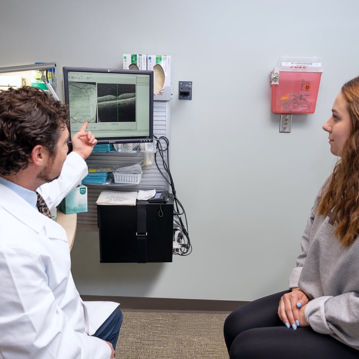
Diagnosing Age-Related Macular Degeneration
The pairing of regular monitoring and annual, comprehensive eye exams offers an effective method to detect AMD and preserve vision. While your specific health needs may facto, men and women 40-54 should visit our retina specialists at least every 2-4 years, or every 1-2 years if over 55. Exams aside, you can monitor your vision on your own at home with the daily use of an Amsler grid (more below).
These exams are the only way ophthalmologists can diagnose dry AMD. You’ll likely undergo dilation, with eye drops widening the pupils (the opening light passes through) to permit a clear, unobstructed retinal view and that of the eye’s back area.
Your doctor can also observe changes and damage to the RPE, which may occur with drusen development. Drusen are small, yellow, cholesterol-like deposits that gather under the retina. While generally not harmful, excess or oversized drusen thin out the macula and make it stop working, causing visual distortions. Retina specialists use drusen size and quantity to assess AMD progression.
If diagnosed with or at risk of developing wet AMD, you may undergo diagnostic testing to identify blood vessel growth, enabling earlier treatment, when it may be more effective. These diagnostic methods may include fluorescein angiography and optical coherence tomography (OCT).
Treatment for Age-Related Macular Degeneration
When it comes to dry AMD, the main treatment is regular monitoring to prevent the condition from progressing into geographic atrophy or wet AMD. Your doctor may also recommend certain vitamins and supplements that have been shown to slow the progression of dry AMD. If geographic atrophy develops, your retina specialist may recommend Syfovre injections, which is currently the only FDA-approved drug available.
For wet AMD, treatment typically depends on symptom severity. Some common treatment options for wet AMD include:
- Anti-vascular endothelial growth factor (anti-VEGF) injections
- Corticosteroid injections
- Focal laser surgery, also known as photocoagulation
- Photodynamic therapy
Age-Related Macular Degeneration FAQs
AMD is very widespread, especially among people 50 years and older. The nation’s main cause of irreversible vision impairment, by 2040, healthcare experts predict almost 288 million cases worldwide. Within the United States, in 2019, it was found that about 20 million people were living with AMD, including 18.34 million with early-stage dry AMD and 1.49 million with late-stage wet AMD. Among those 80 and older, about 3 in 10 have dry AMD, while about 1 in 10 have wet AMD.
Yes, consuming foods containing certain beneficial nutrients has been found to support eye and vision health, and may even delay AMD’s progressive vision loss. Among these foods are dark leafy greens, oily fish, citrus fruits, and nuts. Some people also benefit from taking certain supplements, such as vitamins C and E, and minerals like zinc and copper. If you’re interested in taking supplements for vision health and AMD prevention, talk to your doctor to see if it’s a good option for you.
With dry AMD, daily monitoring of your vision allows for quick observation of any changes. An Amsler grid, a simple paper chart used at home, with horizontal and vertical lines forming a grid pattern, and a dot in the center, offers a simple method. Here are directions to use it:
- Wear your normal reading glasses.
- Make sure it’s located in an area with good lighting.
- Hold the grid about 12-15 inches from your face.
- Cover one eye and focus your uncovered eye on the center dot.
- Check to see if the grid’s lines look straight or distorted, such as wavy lines, blurred or darkened areas, or blank spots.
- Repeat these steps with the other eye.
Alert your ophthalmologist immediately if any changes or distortions appear.
Expert Care for Age-Related Macular Degeneration at Southeastern Retina Associates
Age-related macular degeneration represents a significant threat to vision health, especially among those over 50. At Southeastern Retina Associates, our retina specialist team is uniquely skilled and experienced in diagnosing and treating patients with all forms of macular degeneration, throughout Chattanooga, Knoxville, and the Tri-Cities Region. We encourage you to contact us today to request an AMD consultation.